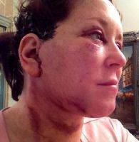
After the facelift procedure, women expect immediate results, but it happens that the face swelling and facelift bruising or hematoma lasts for a long time (up to 2 weeks).
It is worth knowing in advance that postoperative edema is a normal reaction of the body to facelifting. When will it pass? Here everything depends on the individual characteristics of the organism. The process is delayed in the presence of risk factors, which include the patient's age, addiction to smoking, and some medical problems. Blocking the lymph nodes also causes prolonged swelling. In women with thicker skin or overweight, the swelling on the face is more intense.
Photos of bruising after facelift
To reduce the undesirable effects of facelift in Oregon, it is recommended to take some measures. For example, stock up on ice packs and cool your face after the procedure, or put the mattress on the bed at an incline to ensure that the liquid is drained from the face. Avoid too heavy, intense loads, during which there is a profuse sweating. Useful may be a lymphatic massage, which should be used before and after the procedure.
Significantly less edema causes an alternative to classical facelift lifting under local anesthesia. During this procedure, the facial muscles are not cut, but stretched, while reducing the risk of damage to the facial nerves, and the recovery period is much faster. The operation under local anesthesia causes less side effects.
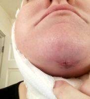
Possible complications after plastic facial skin tightening surgery
Facelift Gone Wrong Photos

The most common complications in performing the braces are related to three main factors:
quality of stopping bleeding, with the reduction of which forms a hematoma;
accuracy of tissue disruption, errors in which lead to nerve damage;
preservation of sufficient blood supply to the flaps formed.
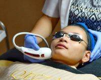
Pros And Cons Of HIFU facelift
Formation of a hematoma. Typical symptoms that indicate the presence of a hematoma under skin-fat grafts are:
the appearance of pain;
an increase in the volume of tissues anterior to the auricle and in the behind-the-ear region (usually on one side);
seepage of fresh blood through the seam line.
Timely diagnosis of this complication is based on constant observation of the patient during the first 4-6 hours after the intervention. With late diagnosis of an increasing hematoma, a skin flap necrosis can develop with catastrophic consequences.
If a strained or growing hematoma is diagnosed, the patient is urgently taken to the operating room. The wound on the corresponding side is opened, blood clots are removed, a source of bleeding is found and stops it. The wound is again sutured with adequate drainage.
Small hematomas, with the condition of stopped bleeding, can be evacuated through the seam line or removed by aspiration. If necessary, repeat the last procedure. Prevention of brusing after facelift formation begins at the stage of preoperative preparation of patients when they are forbidden to take preparations containing acetylsalicylic acid within 3 weeks before the operation, according to indications, treatment aimed at stabilizing the level of arterial pressure is performed. Immediately before the operation, a complete clinical examination is performed. At its final stage, the patient is examined by an anesthesiologist.
Prevention of hematoma during surgery is primarily based on careful stopping bleeding at all sites of the wound. It is performed under conditions of good illumination (using a fiber frontal illuminator) with the help of bipolar coagulation. To improve the hemostatic effect, temporary puffing of the cavities with tissues moistened with 3% hydrogen peroxide solution also helps.
 However, the further actions of the surgeon should be based on an understanding of the obvious fact that with any technique of operation complete certainty in the high quality of stopping bleeding in the wound is not and can not be. The grounds for the illusion of complete hemostasis may arise at the moment preceding the closure of the wound, with careful examination of which there are no signs of bleeding. In this case, the vessels are in an unstable state. After a few tens of minutes, when the action of adrenaline ceases, bleeding from small vessels can resume.
Understanding this mechanism led to the introduction into practice of the method of re-examination of the wound. It consists in the fact that after performing all the manipulations and carefully stopping the bleeding on one side of the face, the wound is swabbed with napkins and not stitched. On the other side of the face, the intervention is carried out to the same stage. After this, the surgeon again returns to the first side and after removing the napkins again examines the wound surface, finally stopping the bleeding. The wound is then sutured and the procedure repeated on the second side.
However, in this case, the guarantees of stopping bleeding are far from absolute. It can resume when the patient is withdrawn from anesthesia, when blood pressure may rise sharply. No less dangerous are the first hours after surgery, when some patients experience nausea and vomiting.
With all this said, the maximum guarantees for the prevention of hematoma formation are the use of drainage tubes with active aspiration of wound contents. With their proper installation under the flaps in the wound, before the moment of its closure, a negative pressure is created, resulting in the wound surfaces being pressed against each other, which helps stop the bleeding from the small vessels after the adrenaline ceases.
Necrosis of the skin develops with insufficient nutrition of the skin flap, which is facilitated by its thinning, excessively extensive detachment of tissues and tension on the seam line. Most often this occurs in the behind-the-ear area, where the line of the greatest tension of the tissues passes. Necrosis of tissues arising in other areas (temporal, pre-prone), as a rule, is a consequence of technical errors.
Suppuration of the wound is a very rare complication, which usually develops when the hematoma is unavailable, as well as due to tissue necrosis. This can be facilitated by the penetration of hair bundles into the wound when seams are applied within the scalp.
Relatively reliable prevention of wound suppuration is achieved in the following ways:
delicate handling of tissues;
preservation of sufficient blood supply to the flaps being formed;
repeated washing of the wound during the operation with solutions of antiseptics;
use (with extensive detachment of tissues) of active drainage systems;
preventive administration of broad-spectrum antibiotics with prolonged interventions.
Hypertrophic scars occur in 1-2% of patients, most often in the behind-the-ear area, where they can be painful. The main reasons for their occurrence are the tension on the seam line and the individual tendency of the patient to form rough scars.
Despite the fact that during the closure of the wound, the tension of the tissues is present only in the zone of the main fixing suture (and is supported by it), in the postoperative period after removal of this seam some tension can be transferred to the bite seam. The direction of this tension can correspond to the seam line. This leads to hypertrophy of the scar.
To prevent the latter allows a change in the configuration of the seam by forming a triangular projection in the hair-free zone. With hypertrophy of the rumen, it is possible to introduce hormones into it. You can apply and the technique of discharge with excision or conservation of the scar.
Deformation of contours. Local changes in the contours of the face after the tightening of the soft tissues are the result of the formation of small hematomas under the skin-fat flaps and (or) the movement of the flaps cut out within the PMPS.
Significant deformations of the contours are, as a rule, the result of inaccurate intersection of platism, inadequate fat removal in the chin area, uneven liposuction and creating an uneven duplication of the edges of the platism along the front line of the neck. These problems are prevented by careful preoperative planning and precise technique of working with tissues. In many cases, the correction of these disorders can be achieved only during the reoperation.
Skin pigmentation. Most often, the pigmentation of visible areas of the skin occurs in patients with sensitive and delicate skin with intradermal hemorrhages. Pigmented spots disappear within a year.
Hair loss occurs in two main forms: local and generalized. With a local form, hair loss occurs in the temporal and occipital areas as a result of the formation of too thin skin flaps, when the surgeon damages the layer in which the hair follicles are located. In most cases, lost hair in the temporal region is restored within 3-4 months. If alopecia in the scar zone is preserved, then it can be eliminated in an operative way: from excising the rumen to rotating the scraps from the scalp.
A generalized form of hair loss occurs under the influence of the stressful situation, which is the patient's surgery. As a rule, this happens in women who had weakness of hair bulbs and a tendency to hair loss before the operation. That is why during the initial examination of patients it is important to pay attention to the condition of the hair and, in the presence of grounds for anxiety, to discuss the possibility of developing this complication. The use of a complex of medicinal products aimed at strengthening hair, as a rule, gives good results.